Programme
-
Seminar #1 - 11 February
Ferroptosis: from metabolism to disease treatment
15:00-15:10
Welcome and Introduction
Mauro Maccarrone
(University of L'Aquila, Italy)15:10-15:40
Fulvio Ursini
(University of Padua, Italy)15:40-16:10
Targeting Ferroptosis for the Treatment of as-yet-Incurable Diseases

16:10-17:00
Questions and Answers
Ferroptosis & Metabolism
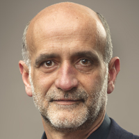
Fulvio Ursini
Dept of Molecular Medicine, University of Padua, ItalyFerroptosis is a form of cell death operated by lipid peroxidation (LPO) under control of GPx4 activity. Since LPO is initiated from traces of LOOH, the question emerges about their formation. The most reasonable candidate is the protonated form of superoxide. While we do not have evidence for a role of respiratory chain, we showed we observed, instead, that it is the pyruvate dehydrogenase complex (PDC) that both produces superoxide and supports ferroptosis when GSH is low. The source of superoxide is the autoxidation of dihydrolipoamide. Further, GSH depletion inhibits the kinase that inhibits PDC. In a perspective of tissue homeostasis, it is the energetic metabolism, associated to a decreased nucleophilic tone, that both supports energy demanding proliferation and sensitizes cells to a regulated form of death.
Fulvio Ursini
Fulvio Ursini, born 1951, serves as professor of biochemistry, School of Medicine, University of Padova. The leitmotif of its studies has been the mechanism of antioxidant effect. These studies led to the discovery of the selenoenzyme GPx4, the functional inactivation of which activates lipid peroxidation and a regulated form of cell death named ferroptosis. He described the chemistry of the functional interaction between enzymatic antioxidant systems-reducing hydroperoxides and chain breaking antioxidants reducing lipid hydroperoxyl radicals. This interplay between a nucleophilic displacement reaction and free radical scavenging contributed to bridging the gap between chemistry and cell biology of free radical oxidations in biology. In the frame of studies on antioxidant mechanisms, he unraveled the role of GPx4 in protein oxidation required for spermatogenesis. This clarified the role of selenium catalysis in male fertility. He first described the post-prandial oxidative stress and the protective role of foods containing nucleophiles (such as wine or beer). The pathophysiological outcome of these studies led to the description of an innovative modification of LDL, where a misfolding of a specific loop of ApoB, where the shift from alpha to beta structure leads to the formation toxic amyloid like aggregates compatible with an atherogenic effect.
Targeting ferroptosis for the treatment of as-yet-incurable diseases
Marcus Conrad
Institute of Metabolism and Cell Death, Helmholtz Zentrum München, Munich, GermanyFerroptosis is a quite recently described metabolic form of non-apoptotic cell death characterized by the iron-dependent oxidative destruction of cellular membranes. Emerging evidence established that ferroptosis is the underlying cause of a number of diseases such as neurodegeneration, tissue ischemia/reperfusion injury and certain cancers. While glutathione peroxidase 4 remains the guardian of ferroptosis, genome-wide CRISPR/Cas and genetic suppressors screens have uncovered additional molecular players that impact on the cells’ susceptibility toward ferroptosis. As such, the ever-growing list of distinct molecular players of the ferroptotic cell death process will aid in the development of target-based novel therapies to alleviate ferroptosis-based diseases.

Marcus Conrad
-
Seminar #2 - 11 March
Redox therapies for neurodegenerative diseases: lights and shadows
15:00-15:10
Welcome and Introduction
Luciano Saso
(Faculty of Pharmacy and Medicine, Sapienza University of Rome)15:10-15:30
The NRF2-KEAP1 Signaling Pathway as a Pharmacological Target in Neurodegenerative diseases
Antonio Cuadrado
(Dept of Biochemistry, Medical School, Autonomous University of Madrid, Spain)15:30-15:50
Breaking the vicious cycle of oxidative stress – inflammation – cell death by reversible cysteine modifications of Keap1

Albena T. Dinkova-Kostova
(Jacqui Wood Cancer Centre, Division of Cellular Medicine, School of Medicine, University of Dundee, UK)15:50-16:10
Oxidative Stress in Amyotrophic Lateral Sclerosis: Pathophysiology and Opportunities for Pharmacological Intervention
Paulo J. Oliveira
(CNC-Center for Neuroscience and Cell Biology, University of Coimbra, UC Biotech Building, Biocant Park, Cantanhede, Portugal)16:10-17:00
Questions and Answers
The NRF2-KEAP1 Signaling Pathway as a Pharmacological Target in Neurodegenerative diseases
Antonio Cuadrado
Dept of Biochemistry, Medical School,
Autonomous University of Madrid, Spain
Email: antonio.cuadrado@uam.esThe etiology of Alzheimer’s disease (AD) remains largely unknown and the therapeutic approaches to stop disease progression have consistently failed. This fact indicates that, in addition to the largely studied amyloid and Tau pathophenotypes, other crucial factors must be considered. Our view is that the loss of homeostatic responses in the elderly is a very relevant factor for AD onset and progression. In recent years, a master regulator of homeostatic responses has been found in the transcription factor NRF2 (Nuclear factor E2-Related Factor 2). NRF2 regulates the expression of over 250 homeostatic genes that play crucial roles in protection against oxidative, inflammatory, metabolic, and proteotoxic forms of cellular stress, all of which are well established pathomechanisms of AD and its co-morbidities. In postmortem brain samples of AD patients, neurons expressing aggregates of TAU or APP exhibit increased levels of the NRF2 targets NQO1 and SQSTM1 and in protein lysates we found upregulation of NRF2 in parallel to increased markers of microgliosis, astrogliosis and inflammation. These results suggest that the increase in NRF2 activity found in the patients is a partially unsuccessful attempt of the diseased brain to compensate the pathologic events. Consistent with this hypothesis, reinforcement of the NRF2 signature in mouse models of AD by daily treatment with an NRF2 activator reduced glial and inflammatory markers and improved cognition and motor complications.
Antonio Cuadrado
Antonio Cuadrado is full professor of Biochemistry and Molecular Biology at the Department of Biochemistry, Medical School, Autonomous University of Madrid. He obtained his PhD degree in 1985 and enjoyed several postdoctoral stays in the National Cancer Institute-NIH with the help of Fulbright and Fogarty fellowships. He established his independent laboratory as Professor of Biochemistry in 1997 with a main interest on the study of molecular mechanisms involved in initiation and progression of neurodegenerative diseases. For the past years his main lane of research has been the validation of transcription factor NRF2, master regulator of cellular homeostasis, with four main lines of activity: i) The transcription factor NRF2 as a new therapeutic target in Parkinson's and Alzheimer’s diseases. ii) Role of oxidative stress in neuronal death and neuroinflammation in neurodegenerative diseases. iii) Pharmacological regulation of autophagy in the brain as a novel therapeutic strategy for neurodegenerative proteinopathies. iv) Development of new NRF2-modulating drugs. Dr. Cuadrado has been awarded several research projects funded by private, local, and governmental agencies to study the molecular basis of Parkinson’s and Alzheimer’s disease. He has published over 150 primary and review articles, of which more than 80 are related to neuroprotection in preclinical models of Parkinson’s and Alzheimer’s disease, that have been cited over 12500 times. He is grant and fellowship evaluation consultant for Spanish governmental and autonomic agencies. As a professor, Dr. Cuadrado has participated in multiple teaching activities for the Bachelor degrees of Biochemistry and Medicine, with special focus on research training. Prof. Cuadrado received a Testimonial of Recognition by the Tohoku Medical Society. Sendai, Japan, in 2014. He is Honorary Scientist at "Victor Babes National Institute of Pahtology" Bucharest, Romania since 2017. In 2019, he was invested Doctor Honoris Causa by Carol Davila University of Medicine and Pharmacy, Bucharest, Romania.
Breaking the vicious cycle of oxidative stress – inflammation – cell death by reversible cysteine modifications of Keap1
Albena T. Dinkova-Kostova
Jacqui Wood Cancer Centre, Division of Cellular Medicine, University of Dundee School of Medicine, Dundee, DD1 9SY, Scotland, United Kingdom
E-mail: a.dinkovakostova@dundee.ac.ukDisrupted redox and protein homeostasis and chronic inflammation engage in a vicious cycle that leads to cell death, and are characteristic features of all neurodegenerative diseases. Transcription factor Nrf2 and its partner Kelch-like ECH-associated protein 1 (Keap1), regulate the expression of large networks of genes encoding proteins that provide powerful and long-lasting protection against damage by oxidants and pro-inflammatory agents. Most pharmacological activators of Nrf2 (termed inducers) are electrophiles that reversibly bind to highly reactive sensor cysteines within Keap1, impairing its repressor function, and allowing for Nrf2 accumulation and enhanced transcription of its target genes. The Keap1/Nrf2 regulatory network includes drug metabolizing, antioxidant, anti-inflammatory, metabolic enzymes, as well as autophagy-related proteins and thus Nrf2 has a critical role in the maintenance of the cellular redox and protein homeostasis, and in the resolution of inflammation. Nrf2 inducers that bind reversibly to Keap1 are protective in cell culture and animal models of neurodegeneration and have documented beneficial effects in humans.
Albena T. Dinkova-Kostova
Albena Dinkova-Kostova graduated in Biochemistry and Microbiology from Sofia University (Bulgaria), and obtained her PhD degree in Biochemistry and Biophysics from Washington State University (USA). She subsequently trained in Pharmacology at Johns Hopkins University School of Medicine (USA), where she continues to hold an Adjunct Professor position. She joined the University of Dundee in 2007 as a Research Councils UK Academic Fellow, and is now Professor of Chemical Biology. Her group collaborates with basic scientists and clinicians and with the pharmaceutical industry. In her research, at the interface of Chemical Biology and Medicine, she is committed to understanding how cells and organisms respond to oxidative, inflammatory, and metabolic stress, and is working towards development of strategies for protection against chronic disease. She is named among the top influential academics in Clarivate’s Highly Cited Researchers 2019 & 2020 lists.
Oxidative Stress in Amyotrophic Lateral Sclerosis: Pathophysiology and Opportunities for Pharmacological Intervention
Paulo J. Oliveira
CNC-Center for Neuroscience and Cell Biology, University of Coimbra, UC Biotech Building, Biocant Park, Cantanhede, Portugal
Email: pauloliv@cnc.uc.ptAmyotrophic lateral sclerosis (ALS), also known as Lou Gehrig's disease or Charcot disease, is a fatal neurodegenerative disease that affects motor neurons (MNs) and leads to death within 2-5 years of diagnosis, without any effective therapy available. Although the pathological mechanisms leading to ALS are still unknown, an excessive reactive oxygen species (ROS) production associated with a weak antioxidant defense may represent an essential pathological feature in ALS. Substantial evidence indicates that oxidative stress (OS) is implicated in the loss of MNs and mitochondrial dysfunction, contributing decisively to neurodegeneration in ALS. Although the modulation of OS represents a promising approach to protect MNs from degeneration, the fact that several antioxidants with beneficial effects in animal models failed to show any therapeutic benefit in patients raises several questions that should be analyzed. We will describe antioxidant compounds tested in ALS preclinical and clinical trials, also describing their respective mechanisms of action. We will describe novel data with mitochondria-targeted dietary polyphenols. While the description of OS mechanism in the different mutations identified in ALS has as principal objective to clarify OS's contribution in ALS, the description of positive and negative outcomes for each antioxidant paves the way for novel opportunities for intervention. Funding: Work in the author's laboratory is funded by the European Regional Development Fund (ERDF), through the COMPETE 2020-Operational Programme for Competitiveness and Internationalisation and Portuguese national funds via FCT–Fundação para a Ciência e a Tecnologia, under projects PTDC/MED-FAR/29391/2017, POCI-01-0145-FEDER-029391 (Mito4ALS), PTDC/BTM-SAL/29297/2017, POCI-01-0145-FEDER-029297 (MitoScreening), and UIDB/04539/2020.
Paulo J. Oliveira
Paulo J. Oliveira is currently Principal Investigator at the CNC – Center for Neuroscience and Cell Biology, University of Coimbra, Portugal. He is the current leader of the “Mitochondria, Metabolism and Disease” group (shorturl.at/rsLRZ) and of the MitoXT: Mitochondrial Toxicology and Experimental Therapeutics laboratory. He received his Bachelor of Science in Biochemistry from the University de Coimbra in 1999. In 2003, he completed his PhD in Cellular Biology from the same University. After completing his doctorate, Paulo Oliveira spent more than three years working at the Medical School of the University of Minnesota, Duluth, USA, where he collaborated with several researchers and contributed to the publication of several peer-reviewed manuscripts. Paulo Oliveira current research interests focus on mitochondrial biology, investigating the alteration of cardiac mitochondrial function by physical activity and diet, cardiac mitochondrial dysfunction and cell death caused by anti-neoplastic agents, mitochondrial alterations during cancer stem cell differentiation and carcinogenesis or rational designing of mitochondria-directed antioxidants. Paulo has about 230 peer-reviewed publications and a large number of collaborations in Europe, Africa and in the USA. He has also received several prizes from the Portuguese Cardiology Society for his work on cardiac mitochondrial research. Besides research, Paulo Oliveira is often involved in teaching at the Department of Life Sciences, University of Coimbra, as well as in other Universities in Portugal and elsewhere, besides in numerous science communication activities. Of importance, he has maintained a consistent primary role in the organization of national and international scientific meetings, including the International Courses in Toxicology (2005-2010 in Coimbra), Annual Meeting of the European Society for Clinical Investigation (2013, Albufeira, Portugal and 2019, Coimbra, Portugal), the 2014 Meeting of the Portuguese Biochemical Society, and FEBS Advanced Lecture Courses in Oncometabolism (2017, Figueira da Foz, Portugal and 2019 Luso, Portugal). Since May 2019, Paulo Oliveira is also President of the European Society for Clinical Investigation, after being its Vice-President for 2 years. Paulo Oliveira has also been reviewer for more than 40 different scientific journals and over 10 different funding agencies, including the European Commission (Research Executive Agency) and the Portuguese Foundation for Science and Technology. His research group is supported by competitive national and international funding agencies, including the Portuguese Foundation for Science and Technology, the European Commission, and private foundations. Paulo is also co-founder of the start-up MitoTAG and is co-inventor in two patents. Since 2020, Paulo is Vice-President of the CNC – Center for Neuroscience and Cell Biology, University of Coimbra, Portugal.
-
Seminar #3 - 26 April
Crosstalk between oxidative stress and metabolic defects: implications for Alzheimer's disease
15:00-15:10
Welcome and Introduction
Luciano Saso
(Faculty of Pharmacy and Medicine, Sapienza University of Rome)15:10-15:40
Amyloid β-peptide and brain oxidative damage: focus on the intersection of the lipid peroxidation product HNE covalently bound to brain proteins, glucose dysmetabolism, and Alzheimer disease

D. Allan Butterfield
(Dept of Chemistry and Sanders-Brown Center on Aging, University of Kentucky, Lexington, KY, USA)15:40-16:00
Mitochondrial dysfunction and oxidative stress in Alzheimer’s disease

Paula I. Moreira
(Institute of Physiology, Faculty of Medicine, Center for Neuroscience and Cell Biology, University of Coimbra, Portugal)16:00-16:20
Down Syndrome is a metabolic disease: altered insulin signaling mediates brain dysfunctions

Marzia Perluigi
(Dept of Biochemical Sciences, Sapienza University of Rome, Italy)16:20-17:00
Questions and Answers
Amyloid β-peptide and brain oxidative damage: focus on the intersection of the lipid peroxidation product HNE covalently bound to brain proteins, glucose dysmetabolism, and Alzheimer disease
D. Allan Butterfield
Dept of Chemistry and Sanders-Brown Center on Aging
University of Kentucky, Lexington, KY, USAAlzheimer disease (AD), an age-related neurodegenerative disorder, is characterized pathologically by the presence of senile plaques (rich in amyloid beta-peptide (1-42) [Aβ(1-42)], neurofibrillary tangles (hyperphosphorylated tau), and neurite and synapse loss, and clinically by progressive diminution of memory and loss of higher executive cognitive functions, resulting in dementia. Knowledge of the underlying etiology and pathogenesis of AD is needed to effectively use agents to retard or prevent this ongoing and worsening public health crisis. Oligomeric Aβ(1-42) is neurotoxic and associated with oxidative damage in vitro and in vivo. Protein oxidation is indexed by elevated protein carbonyls and 3-nitrotyrosine, while lipid peroxidation is indexed in our laboratory by elevation of protein-bound 4-hydroxynonenal (HNE). Redox proteomics, pioneered in the Butterfield laboratory, identified oxidatively dysfunctional brain proteins in oligomeric Aβ(1-42) preclinical models and human studies, including amnestic mild cognitive impairment (MCI), and familial-, preclinical-, early-, and late-AD, Down syndrome (DS), and DS with AD (the DS studies in collaboration with Prof. Marzia Perluigi, Sapienza University of Rome). Among the major pathways affected by Aβ(1-42)-associated lipid peroxidation and consequent HNE bound to proteins are those involved in glucose metabolism, known to decreased in AD and MCI brains. That is, Ab42-mediated lipid peroxidation, leading to HNE and protein oxidative modification and dysfunction, occurs early in AD pathogenesis, resulting in decreased glucose metabolism, including diminished glycolysis, TCA cycle efficiency, and mitochondrial function with consequent loss of ATP production. This, in turn, leads to excitotoxicity, loss of neurotransmission, synaptic plasticity and cell structure, elevated neuroinflammation, impaired protein folding and degradation, and altered signal transduction. We propose oligomeric Aβ(1-42)-associated oxidative modification of brain glycolytic and TCA enzymes and ATP synthase in AD and MCI brains leads to glucose dysmetabolism, resulting in decreased ATP production with consequent loss of neuronal cell potentials and large influx of neurotoxic Ca2+. In turn, prolonged elevated Ca2+ causes necrotic and apoptotic neuronal death indexed by decreased thickness of hippocampus (memory loss) and frontal cortex (higher executive function loss). Recent neuroimaging studies on three continents involving thousands of pre-symptomatic persons carrying genes that cause familial AD for over 22 years prior to onset of symptoms and three years thereafter precisely confirmed the above order of events in AD brains proposed by our laboratory. Consistent with our hypothesis, we demonstrated that synaptic membranes, isolated from brains of targeted-replacement mice in which human apolipoprotein E2, E3, or E4 replaced mice ApoE, when treated with Aβ(1-42) led to elevated oxidative damage in the order apoE4 > apoE3 > apoE2. This is the same order of elevated risk of developing AD as a function of ApoE allele. We hypothesize that apoE4, which lacks key Cys residues and therefore does not bind HNE, in marked contrast to efficient binding of HNE by one Cys residue in apoE3 and two Cys residues in apoE2, does not prevent binding of neurotoxic HNE to key glucose metabolic proteins. Thereby, apoE4 contributes to the elevated risk of developing AD by those who inherit the ApoE4 allele. Our studies identified potential therapeutic targets, which if protected by pharmacology, lifestyle interventions or treatments to reduce Aβ production or limit HNE-mediated damage caused by lipid peroxidation induced by this neurotoxic peptide, could be beneficial in preventing or delaying the progression of this devastating dementing disorder. Support: NIH grants to Professor Butterfield.
D. Allan Butterfield
Professor Butterfield earned his Ph.D. in Physical Chemistry from Duke University (Durham, North Carolina), followed by a National Institutes of Health (NIH) Postdoctoral Fellowship in Neurosciences at the Duke University School of Medicine. In 1975, he joined the Faculty of the Department of Chemistry at the University of Kentucky and quickly rose through the ranks to Full Professor. He is currently the University of Kentucky Alumni Association Endowed Professor of Biological Chemistry, Associate Vice President for Research, Director of the Redox Metabolism Core of the National Cancer Institute-designated UK Markey Cancer Center, and among the Faculty of the world-renowned Sanders-Brown Center on Aging. Professor Butterfield’s research centers on oxidative damage, redox proteomics, and alterations in glucose metabolism and autophagy (and other aspects of the proteostasis network) in brains of persons with Alzheimer disease and Down syndrome, as well as brain alterations in cancer survivors who demonstrate cognitive loss following chemotherapy. Prof. Butterfield has trained 67 Ph.D. and M.S. students and 30 postdoctoral scholars (5 of whom are from the Sapienza University of Rome), which has led to his more than 670 scientific publications [h-index = 112]. His research has been continuously funded for 46 years by NIH or other federal and private funding agencies. Professor Butterfield was recognized recently as the 10th most important scientist in the world in the field of Alzheimer disease research (of approximately 150,000 AD researchers) for the period 2010-2020 based on data mining of the PubMed website. He received the Presidential Award for Excellence in Science Mentoring from President Clinton in the White House, and Prof. Butterfield was elected a Fellow of the Society for Redox Biology and Medicine, an international scientific society from whom he also was presented the 2013 Discovery Award for his seminal and paradigm-shifting research on the role of oxidative stress in the etiology and pathogenesis of Alzheimer disease associated with amyloid beta-peptide.
Mitochondrial dysfunction and oxidative stress in Alzheimer’s disease
Paula I. Moreira
CNC – Center for Neuroscience and Cell Biology
University of Coimbra & Faculty of Medicine
University of Coimbra, Coimbra, PortugalMitochondria fulfill a number of essential cellular functions, and it is recognized that the strict regulation of the structure, function, turnover and transport of these organelles is a control node for the maintenance of cellular homeostasis. Although the precise culprit in the pathogenesis of Alzheimer’s disease (AD) is still obscure, mitochondria and redox imbalance have been proposed to be early events. Studies from our laboratory demonstrate that mitochondria from AD preclinical models are characterized by a defective respiratory chain and an impaired phosphorylation system culminating in an energy crisis. An increased production of mitochondrial reactive oxygen species, reduced antioxidant defenses and defective turnover were also observed. Interestingly, our studies also demonstrate that AD shares several common features with type 2 diabetes including a similar profile of mitochondrial abnormalities and oxidative stress. Our findings suggest that strategies aimed at ameliorating mitochondria and redox status could represent effective preventive and/or therapeutic strategies.
Paula I. Moreira
Paula I. Moreira received her PhD in Biomedical Sciences from University of Coimbra, Portugal. She is Assistant Professor at Faculty of Medicine, University of Coimbra and Principal Investigator at the Center for Neuroscience and Cell Biology, University of Coimbra, Portugal. Moreira’s research team is interested in elucidating the role of mitochondria, metabolic hormones and sex dimorphism in brain changes occurring in diabetes and age-related neurodegenerative diseases, particularly Alzheimer’s disease. Pharmacological and non-pharmacological strategies capable of ameliorating mitochondria homeostasis are also being investigated as possible treatments. Moreira is author of > 200 peer-reviewed publications (H-index = 62; > 19500 citations; Scopus) and serves on the editorial board of several international peer-reviewed journals including Antioxidants & Redox Signaling, Biochimica et Biophysica Acta – Molecular Basis of Disease and Journal of Alzheimer’s Disease. She was a member of the Committee on Novo Nordisk Foundation Challenge Programme 2020 – Biomedicine and Health Sciences and is an ad hoc reviewer for Marie Sklodowska-Curie Individual Fellowships, European Science Foundation and Alzheimer’s Association, among others. Moreira won the Stimulus to Research prize, in 2003, supported by the Calouste Gulbenkian Foundation and, in 2008, the L’Oréal for Women in Science Award supported by L'Oreal Portugal/UNESCO/FCT.
Down Syndrome is a metabolic disease: altered insulin signaling mediates brain dysfunctions
Marzia Perluigi
Dept of Biochemical Sciences
Sapienza University of Rome, ItalyDown Syndrome (DS) is the most frequent chromosomal abnormality that causes intellectual disability, resulting from the presence of an extra complete or segment of chromosome 21 (HSA21). In addition, trisomy of HSA21 contributes to altered energy metabolism that appears to be a strong determinant in the development of pathological phenotypes associated with DS. Alterations include, among others, mitochondrial defects, increased oxidative stress levels, impaired glucose, and lipid metabolism, finally resulting in reduced energy production and cellular dysfunctions. These molecular defects seem to account for a high incidence of metabolic disorders, i.e., diabetes and/or obesity, as well as a higher risk of developing Alzheimer's disease (AD) in DS. A dysregulation of the insulin signaling with reduced downstream pathways represents a common pathophysiological aspect in the development of both peripheral and central alterations leading to diabetes/obesity and AD. Considering that DS subjects are at high risk to develop early onset Alzheimer disease, they can offer a unique opportunity to decipher the link between trisomy of HSA21 and defects of insulin and insulin-related pathways in both aging and neurodegeneration. Drawing the molecular signature underlying these processes in DS is a key challenge to identify novel drug targets and set up new prevention strategies aimed to reduce the impact of AD in DS.

Marzia Perluigi
Marzia Perluigi’s laboratory focused the attention on the analysis of oxidative modifications of proteins and how dysfunction of selected proteins translate into pathological features of a disease state. By following this approach we contributed to shed light on critical molecular determinants underlying aging and cognitive dysfunction. Particularly, my team focuses on redox mechanisms of neurodegeneration in Down Syndrome (DS) and Alzheimer Disease (AD). Results obtained by the analysis of human specimens and studies from mouse and cellular models of the disease reveal a molecular link between protein oxidation/aggregation, the integrity of the protein quality control system (proteasome, UPS and autophagy), dysfunction of energy metabolism and neurodegeneration. Many common pathological hallmarks exist between DS and AD, including deposition of amyloid plaques, NFTs, increased oxidative damage and impaired mitochondrial function, among others. Intriguingly, we propose that all of these processes seem to be joined by a ‘leitmotif’ – oxidative stress – since they are all the cause and/or the consequence of increased free-radical burden. In addition, other than proteostasis and glucose metabolism, redox proteomics studies allowed the identification of oxidized proteins belonging to several dysfunctional pathways among which, detoxification systems, excitotoxicity or synapse function that highly correlates with DS and AD pathological features supporting the role of protein oxidative damage in neurodegeneration and cognitive decline. Further, we recently demonstrated the disturbance of PI3K/Akt/mTOR axis in DS brain, prior and after development of AD. Aberrant mTOR signalling in the brain affects multiple pathways including glucose metabolism, energy production, mitochondrial function and autophagy. All these events are key players in age-related cognitive decline and contribute to the development of Alzheimer-like dementia in DS. Restoration of mTOR signaling may be a powerful strategy to prevent/delay the onset of dementia in DS as well as in the general population. She is currently the Chair of European Chapter of the T21Research Society.
-
Seminar #4 - 13 May
Radioprotectors and radiosensitizers in cancer therapy
15:00-15:10
Welcome and Introduction
Luciano Saso
(Faculty of Pharmacy and Medicine, Sapienza University of Rome)15:10-15:30
Targeting redox metabolism for cancer therapy: from the bench to the bedside
Douglas R. Spitz
(Department of Radiation Oncology, Holden Comprehensive Cancer Center, The University of Iowa, Iowa City, IA, USA)15:30-15:50
SOD mimics act as a radiosensitizer by enhancing hydrogen peroxide levels and altering metabolism in prostate cancer cells

Rebecca Oberley-Deegan
(Department of Biochemistry and Molecular Biology, University of Nebraska Medical Center, Omaha, NE, USA)15:50-16:10
Advancing redox principles and chemistry for diagnosis and treatment of radiation resistant cancer
Cristina M. Furdui
(Department of Internal Medicine, and Co-Director of the Center for Redox Biology and Medicine at Wake Forest School of Medicine, Winston-Salem, NC, USA)16:10-17:00
Questions and Answers
Targeting Redox Metabolism for Cancer Therapy:
From the Bench to the BedsideDouglas R. Spitz, PhD
Free Radical and Radiation Biology Program, Department of Radiation Oncology, Holden Comprehensive Cancer Center, The University of Iowa, IA, USARelative to normal cells, cancer cells have been shown to demonstrate fundamental alterations in oxidative metabolism leading to increased steady-state levels of superoxide, hydrogen peroxide, and labile iron pools. Alterations in cancer cell oxidative metabolism in turn lead to disruptions in many redox sensitive signaling and gene expression pathways that impact cell growth, differentiation, genomic instability, and cancer progression. Cancer cells adapt to this altered metabolic state by activating many signaling and gene expression pathways that allow them to survive and continue to progress to malignancy. The current talk will discuss how pharmacological ascorbate and superoxide dismutase mimics can be used to target the oxidative metabolic frailties of cancer versus normal cells to enhance therapeutic outcomes to radio-chemo-therapies both in pre-clinical and clinical trials. The long-term goal is to utilize a detailed mechanistic understanding of alterations in cancer versus normal cell oxidative metabolism and redox biology to develop rapidly implementable new strategies that can enhance tumor responses while protecting normal tissues during standard radio-chemo-therapy protocols. (supported by P01 CA217797, P01 CA244091, P50 CA174521, P30 CA086862, R01 CA182804, R50 CA243693, T32 CA078586)
Douglas R. Spitz
Douglas R. Spitz, PhD is a Professor of Radiation Oncology, Director of the Free Radical and Radiation Biology PhD Program, and Director of the Free Radical Metabolism and Imaging Program in the Holden Comprehensive Cancer Center at the University of Iowa. Dr. Spitz received his B.A. in 1978 from Grinnell College and his Ph.D. in 1984 from The University of Iowa working with Dr. Larry Oberley. He did his post-doctoral work at the University of California San Francisco with Drs. Gloria Li and William Dewey and has held faculty positions at the University of Virginia, Washington University in St. Louis, and the University of Iowa. His research is supported by the NIH/NCI to participate in both basic science and translational cancer research projects designed to target oxidative metabolism for improving responses to cancer therapy.
SOD mimics act as a radiosensitizer by enhancing hydrogen peroxide levels and altering metabolism in prostate cancer cells
Rebecca Oberley-Deegan
Department of Biochemistry and Molecular Biology, University of Nebraska Medical Center, NE, USAProstate cancer patients are often treated with radiotherapy. MnTE-2-PyP, is a superoxide dismutase (SOD) mimic and a known radioprotector of normal tissues. Our recent work demonstrates that MnTE-2-PyP also inhibits prostate cancer progression with radiotherapy; however, the mechanisms remain unclear. We have identified that MnTE-2-PyP-induced intracellular H2O2 levels are critical in inhibiting growth of prostate cancer cells. High H2O2 levels by MnTE-2-PyP treatment induced nuclear fragmentation in PC3 cells, which could be synergistically enhanced with radiotherapy both in vitro and in vivo. In LNCaP cells, disturbing H2O2 balance may contribute to the bi-nucleation phenomenon. We found that MnTE-2-PyP induced protein oxidations in PC3 cells and one major group of oxidized protein targets were involved in energy metabolism. The oxidative phosphorylation rates were significantly enhanced in both PC3 and LNCaP cells with MnTE-2-PyP treatment, but mitochondrial membrane potential was unaffected. In addition, MnTE-2-PyP significantly increased NAD(P)+/NAD(P)H ratios in PC3 and LNCaP cells in a dose-dependent manner, which was mainly due to a reduction of cellular NAD(P)H pool. Correspondingly, we observed a significant decrease of activity in glucose-6-phosphate dehydrogenase (G6PD) and 6-phosphogluconate dehydrogenase (6PGD), which are major cellular NADPH producing enzymes in pentose phosphate pathway. A decrease of GSH/GSSG ratios were confirmed in MnTE-2-PyP-treated prostate cancer cells, which may result from the decreased glutathione reductase (GR) activity due to NADPH depletion. We conclude that the increased H2O2 levels, protein oxidative modifications, mitotic catastrophe, cellular energy metabolism alterations, and NAD(P)H depletion caused by MnTE-2-PyP are all likely factors contributing to prostate cancer cell growth inhibition.
Rebecca Oberley-Deegan
Dr. Rebecca Oberley-Deegan received her Ph.D. University of Iowa, Iowa City Iowa and did her postdoctoral training at National Jewish Health, Denver, Colorado. She joined the University of Nebraska Medical Center (UNMC) in Omaha, Nebraska in 2014 as an assistant professor and is currently an associate professor at UNMC. Dr. Oberley-Deegan’s laboratory has previously shown that a catalytically active antioxidant can protect normal prostate tissues during radiation but not prostate tumor tissues. The focus of her research is determining the mechanisms by which antioxidants can protect normal tissues from radiation while simultaneously making the tumor vulnerable to radiation damage. Dr. Oberley-Deegan’s is focusing specifically on the role of free radical signaling to transform fibroblasts and inflammatory cells in normal tissues, which results in damage to these tissues. In the context of prostate cancer, her group is focusing on inhibiting the signaling events controlled by radiation-induced free radicals involved in tumor survival and metastasis.
Advancing Redox Principles and Chemistry for Diagnosis and Treatment of Radiation Resistant Cancer
Cristina M. Furdui
Department of Internal Medicine, Section on Molecular Medicine, Wake Forest School of Medicine, NC, USARedox metabolism is a driving regulator of cancer progression, response to therapies and long-term patient’s quality of life. Well-established cancer therapies, including radiotherapy, either directly impact redox metabolism or have redox-dependent mechanisms of action defining their clinical efficacy. This seminar will present an overview of principles, reagents and methods for tracking redox processes in biological systems focusing on protein sulfenylation, and how these are applied in conjunction with computational modeling to gain knowledge of the mechanisms underlying the resistance to radiotherapy in Head and Neck Cancer (HNC). Furthermore, the seminar will present new applications of protein sulfenylation reagents as chemotherapeutics and PET imaging diagnostics for radiation resistant HNC.
Cristina M. Furdui
Dr. Cristina Furdui is Professor in the Department of Internal Medicine, Section on Molecular Medicine with joint appointments in the Departments of Biochemistry and Cancer Biology at Wake Forest School of Medicine, and affiliate faculty in the Department of Chemistry, Wake Forest University. She serves as Co-Director of the Center for Redox Biology and Medicine and the NIH T32 training program in Redox Biology and Medicine, and Director of the Proteomics and Metabolomics Shared Resource supported by the Wake Forest Comprehensive Cancer Center. She chaired the Gordon Research Conference on Thiol-Based Redox Regulation & Signaling in 2016 and serves on the Editorial Board for J Biol Chem, Antioxidants, and Chem Res Toxicol. Her research combines chemical and systems biology approaches to interrogate redox mechanisms of disease. Current projects focus on: 1) development of chemical probes and methods for analysis of protein oxidation; 2) functional studies of redox-regulated proteins; 3) cancer prevention, treatment and quality of life with emphasis on lung and head and neck cancer; 4) mechanisms and new therapies targeting inflammation and sepsis; and, 5) biospecimen preservation to advance analysis of clinical biomarkers and precision medicine. Dr. Furdui is also the co-founder of Xoder Technologies, LLC, a company dedicated to the advancement of redox knowledge.
-
Seminar #5 - 24 June
Therapeutic potential of antioxidants: from male infertility to retinal disorders
15:00-15:10
Welcome and Introduction
Mauro Maccarrone
(University of L'Aquila, Italy)15:10-15:40
Oxidative stress and antioxidant-based therapies in male infertility

Barbara Tavazzi
(Catholic University of Rome, Italy)15:40-16:10
A glance on antioxidants: the role of sulfur amino acids in retinal disorders
Marcello Allegretti
(Dompè, L’Aquila, Italy)16:10-17:00
Questions and Answers
Oxidative stress and antioxidant-based therapies in male infertility
Barbara Tavazzi
Catholic University of Rome, ItalyUnder physiological conditions, reactive oxygen species (ROS) play pivotal roles in various processes of human spermatozoa. Indeed, semen requires the intervention of ROS to accomplish different stages of its maturation. Overproduction of ROS is potentially harmful, and may concomitantly occur to excess generation of reactive nitrogen species (RNS), leading to oxidative/nitrosative stress. Spermatozoa are provided with a set of scavenger enzymes and human seminal plasma has a specific pattern of water- and fat-soluble low-molecular weight antioxidants aimed to decrease risks associated with oxidative/nitrosative stress. Various studies clearly demonstrated that male infertility is characterized by various signs of molecular damages caused by increased ROS and RNS production. Therefore, clinical studies have been conducted using the administration of antioxidant-based therapies as potentially valid tool to treat male infertility.
Barbara Tavazzi
Prof. Barbara Tavazzi received her Ph.D. University of Rome La Sapienza, and did her postdoctoral training at University of Rome "Tor Vergata", Rome, Italy where she remain as researcher until to 2003. Starting from that year at present, she joined the Catholic University of Rome as Associate Professor in Biochemistry and has assistantship at the Diagnostic Area and Infectious Diseases, Clinical Chemistry, Fondazione Policlinico Universitario A. Gemelli IRCCS of Rome. In the October 2021, she will start her activity as Full Professor of Biochemistry at the Faculty of Medicine and Surgery of the Saint Camillus International University of Health and Medical Sciences (UniCamillus) in Rome.
The focus of her research is oriented mainly towards research areas related to biochemistry, clinical biochemistry and translational medicine, in particular the field of metabolomics, targeted proteomics and regulation and gene expression. The main lines of research can be summarized as follows:
biochemical assessment (energy metabolism, mitochondrial function, oxidative/nitrosative stress, enzymatic activities); biochemical (energy metabolism, mitochondrial function, oxidative stress/nitrosis, enzyme activities) and molecular (gene expression) assessment of brain damage in experimental head trauma (weight drop model with diffuse axonal damage); analysis of low molecular weight metabolites in biological fluids (CSF, blood, urine) for the assessment of oxidative/nitrosative stress damage and alterations of energy metabolism in human head trauma; research of circulating and urinary biomarkers (metabolic, protein and molecular) with prognostic value in chronic neurodegenerative diseases (multiple sclerosis, amyotrophic lateral sclerosis); research on circulating and urinary biomarkers (metabolic, protein and molecular) in the pathology of male and female infertility, with particular attention to metabolomic parameters and oxidative and nitrosative stress.A glance on antioxidants: the role of sulfur amino acids in retinal disorders
Marcello Allegretti, PhD
Chief Scientific Officer Dompé farmaceutici S.p.A., ItalyOxidative stress plays a major role in the neurodegenerative process. Retinal cell survival involves a balance between reactive oxygen species (ROS) and antioxidant scavengers to counteract oxidative stress (OS) injury. The retina is susceptible to OS due to its elevated oxygen consumption and exposure to visible light, which results in cellular damage caused by ROS. Elevated OS levels determine dramatic changes that lead to visual impairment. Age-related macular degeneration (AMD), diabetic retinopathy (DR), and glaucoma are ocular disorders that can lead to visual loss, and for which the involvement of ROS has been evoked. The retina is endowed with an efficient innate immune system that activates three essential pathways: migration of microglia, stimulation of the complement system, and inflammasome assembly in the retinal pigment epithelium (RPE). For this response, retinal cells are endowed with a variety of immune receptors and mediators in charge to help the cells to eliminate the insult. Under OS, the activation of this immune pathway aims to repair tissue homeostasis. Still, under continual stress, the inflammatory system's chronic hyperactivation can determine dramatic tissue changes and damage, resulting in irreversible retinal pathologies, including AMD or DR.
Sulfur amino acids (SAAs), such as methionine and taurine are necessary for the synthesis of proteins and serve as precursors of important cofactors and metabolites, thus playing a critical role in health maintenance. Methionine and taurine are present in ocular tissues, such as cornea, iris, lens, ciliary bodies, vitreous, and retina. Although their role in the retina is unclear, numerous events have been attributed to them, involving osmoregulation, antioxidant defence, stress responses, and protein stabilization.
The role of sulfur amino acids as regulators of retinal cell functions will be discussed.Marcello Allegretti
Since 2009:
- Chief Scientific Officer at Dompé farmaceutici managing Dompé’s Research and Development activities and pre-clinical and clinical drug development programs - from active ingredient identification to market authorization - including relationships with international regulatory authorities.
- Number of university teaching positions.
- Published work in over 40 scientific journals.
- Over 30 international patents.
Prior 2009:
- Degree in Medical Chemistry from The Sapienza University of Rome.
- Member of the American Chemical Society.
- Member of the Italian Chemistry Society.
-
Seminar #6 - 6 July
Interplay between the gaseous signaling molecules NO and H2S: from reaction mechanisms to physiology
15:00-15:10
Welcome and Introduction
Alessandro Giuffrè
(Consiglio Nazionale delle Ricerche, Italy)15:10-15:40
The reactive species interactome in health and disease

Martin Feelisch
(University of Southampton, United Kingdom)15:40-16:05
The H2S/NO crosstalk at the level of the H2S-synthesizing enzymes
João B. Vicente
(Universidade Nova de Lisboa, Portugal)16:05-16:30
The redox physiology and pharmacology of the interaction of sulfide and nitric oxide in the cardiovascular system

Miriam Cortese-Krott
(University of Dusseldorf, Germany)16:30-17:00
Questions and Answers
The H2S/NO crosstalk at the level of the H2S-synthesizing enzymes
Joao B. Vicente
Universidade Nova de Lisboa, PortugalIn mammalian physiology, the so called ‘gasotransmitters’ nitric oxide (NO), carbon monoxide (CO) and hydrogen sulfide (H2S) are reactive gaseous molecules synthesized endogenously to accomplish numerous signaling roles. Sharing similar mechanisms of action, namely by interaction and/or modification of proteins, gasotransmitters have been shown to regulate numerous cellular functions and physiological processes. Accordingly, disturbed levels of each gasotransmitter due to aberrant expression of enzymes involved in their synthesis or breakdown have been associated with multiple human diseases, including cardiovascular, neurodegenerative diseases and cancer.
Interestingly, gasotransmitters exhibit a notable degree of crosstalk, where the bioavailability of one controls that of the others. Herein, I will present an overview of investigations on the control of the H2S synthesizing enzymes cystathionine β-synthase and cystathionine γ-lyase by NO and NO-related species. This crosstalk entails numerous their (patho)physiological implications and offers opportunities for pharmacological interventions.João B. Vicente
João B. Vicente earned his European Label PhD in Biochemistry from NOVA University of Lisbon (Portugal) and Sapienza University of Rome (Italy) in 2007. After a post-doc in Stanford University (USA) and a researcher position at the School of Pharmacy, Lisbon University (Portugal), he started a position as Auxiliary Investigator in the Macromolecular Crystallography Unit at NOVA Instituto de Tecnologia Química e Biológica (Portugal). His research team combines protein biochemistry and biophysics with structural biology to study the molecular mechanisms underlying human hydrogen sulfide (H2S) metabolism. Particularly, his team is interested in understanding how the metabolic pathways leading to the production and breakdown of H2S are regulated by endogenous physiologically relevant modulators. Through a network of collaborators, he aims to position these metabolic pathways and their regulatory mechanisms as key determinants in the context of various human diseases. Moreover, he is committed to the discovery and development of pharmacological and diagnostics tools to improve therapeutics.
The redox physiology and pharmacology of the interaction of sulfide and nitric oxide in the cardiovascular system
Miriam Cortese-Krott, PhD
University of Dusseldorf, GermanyThe interest in the biological chemistry of the chemical interaction of nitrogen-bearing and sulfur-bearing compounds was recently revived by the discovery that NO and sulfide may react with each other leading to the formation of “S-N hybrid species” with highly specific biological and pharmacological activity. These include nitrosopersulfide (SSNO−), polysulfides (HSn-), and dinitrososulfite [N-nitrosohydroxylamine-N-sulfonate, a NONOate derivative of sulfite]. Each of these products is characterized by distinct chemical biology and in vitro and in vivo bioactivity, and their generation is accompanied by the formation of several N- and S-oxides including thiosulfate and nitroxyl (HNO). Polysulfides appear to play a key role in both, generation and degradation of SSNO-. This webinar will provide a brief overview of the cardiovascular effects of each of the three main products along with a summary of their main pathways of formation and decomposition. Our results suggest that wherever NO and sulfide are co-generated in a biological environment, their bioactivity is governed by an unexpectedly rich network of reactive species and intermediates, like polysulfides and anionic S/N-hybrid species. The availability of precursors and targets of the reactive species generated by these interactions may contribute to modulate the redox physiology of the cardiovascular system.
Miriam Cortese-Krott
Miriam M. Cortese-Krott graduated with honors in Pharmaceutical Biotechnology from the Faculty of Pharmacy, University of Milan, Italy, in 2002. Her thesis was focused on transgenic mouse models of atherosclerosis. She received her PhD with honors in Pharmacology from the University of Düsseldorf, Germany in 2007 with a thesis on the role of nitric oxide in inflammation and atherosclerosis. After a post-doc focused on stem cell redox signaling in the Department of Surgery of the RWTH Aachen University, Germany, she moved back to Düsseldorf for a second postdoc in the Department of Cardiology, Pneumology, and Angiology, University Hospital, where she isolated and characterize nitric oxide metabolism in red blood cells and its downstream pathway, and created tissue specific eNOS knockout and knockin mouse models. She obtained her Habilitation in 2014 in Molecular Medicine. Since 2016 she was appointed a tenured professorship in myocardial infarction research, and since 2019 she is the head of the myocardial infarction research laboratory.
Martin Feelisch
After the study of pharmacy, Martin Feelisch earned his PhD in Pharmacology from Heinrich-Heine University in Dusseldorf and a habilitation (postdoctoral research and teaching qualification) in Pharmacology and Toxicology at the University of Cologne. He was appointed to the Personal Chair of Experimental Medicine & Integrative Biology at the University of Southampton in 2012, following a series of senior research positions in both industry (Director of Pharmacology, Schwarz Pharma) and academia (University College London, Louisiana State University Health Sciences Center, Boston University School of Medicine, Warwick University) in the US and in Europe.
Martin is a Systems Biologist and Analytical Chemist with particular interests in cardiovascular pharmacology, intermediary metabolism, evolutionary biology and photobiology, disease mechanisms, and the chemical biology of nitric oxide, nitroxyl and sulfur in its various forms. More recent research areas include the role of sunlight for cardiovascular health, mechanisms of adaptation to hypoxia and other stressors, and the significance of nutrition for health and resilience. His particular interests in the chemical interactions between reactive oxygen, nitrogen and sulfur species (ROS, RNS, RSS) related to mitochondrial function, redox signalling and electron balance at the whole-body level led to the development of the ‘Reactive Species Interactome’ concept and a conceptual framework known as the ‘Redox Interactome’.
Work in Martin’s lab is both hypothesis- and curiosity-driven and includes basic as well as translational research projects in all of the above areas. Current development efforts in his lab focus on targeted metabolomic approaches (‘redox metabolomics’) to disentangle mechanisms of redox regulation in health and disease. -
Seminar #7 - 7 September
Redox balance, metabolism and aggressiveness of cancer cells
15:00-15:10
Welcome and Introduction
Mauro Maccarrone
(University of L'Aquila, Italy)15:10-15:40
Mitochondrial redox hubs as promising targets for anticancer therapy

Paola Chiarugi
(University of Florence, Italy)15:40-16:10
Targeting NF-κB downstream effectors in cancer: embracing complexity to treat disease

Daria Capece
(University of L’Aquila, Italy)16:10-17:00
Questions and Answers
Mitochondrial Redox Hubs as Promising Targets for Anticancer Therapy
Paola Chiarugi
University of Florence, ItalyBeyond their bioenergetic role, mitochondria are essential for providing malignant cells a higher plasticity to face the harsh environmental conditions. Metabolic adaptation to microenvironmental cues represents the triggering event of mitochondria overexploitation to orchestrate nutrient sensing and upload, signaling, and redox circuits. Mitochondria produce high amounts of ROS functional for multiple signaling networks underlying tumor proliferation, survival, and metastatic process. Key molecular pathways are regulated through mitochondrial ROS, including HIF-1 stabilization, SIRT1/3 activity, Src kinase and recptor tyrosine kinase activity, with a consequent impact on hallmark features of cancer aggressiveness as cell adhesion, motility, epithelial mesenchymal transition and environmental/pharmacological stress survival.
Paola Chiarugi
Paola Chiarugi is Full Professor of Biochemistry in the Faculty of Medicine and Surgery at the University of Florence. She is member of the Excellence and Research Centre for Transfer and High Formation: Studies at Molecular and Clinical Level of Chronic, Inflammatory, Degenerative and Neoplastic Diseases, of the Faculty of 1000 Evaluation System and of Italian Society for Biochemistry and Molecular Biology.
Paola Chiarugi has studied for up to 10 years the structure-function relationship of tyrosine phosphatises, contributing to the elucidation of the mechanism of action of these enzymes and to the definition of their role in the control of cell proliferation, adhesion and motility. More recently her interests moved toward the redox regulation of oxidant-sensitive proteins during cell proliferation and cell adhesion to extracellular matrix, particularly focusing on the role of tyrosine phosphorylation on chemo-attractant and chemo-repulsive receptor signalling as ephrins and Eph kinase receptors. Actually she is investigating the role of the redox-regulated molecular pathways affecting metastatic conversion of cancer cells, particularly focusing on oxidative stress and hypoxia as key determinants for epithelial-mesenchymal or mesenchymal-amoeboid transition of invasive tumour cells.Targeting NF-κB downstream effectors in cancer: embracing complexity to treat disease
Daria Capece
University of L’Aquila, ItalyNF-κB transcription factors drive oncogenesis, disease recurrence and therapy resistance in most human cancers. As such, NF-κB is a potential therapeutic target in many tumours. However, the severe on-target toxicities associated with systemic NF-κB inhibition have thus far precluded the development of specific, clinically useful, NF-κB inhibitors. We reasoned that an attractive alternative to safely target NF-κB would be to block the non-redundant, cancer-specific effectors of NF-κB-dependent functions. To this end, we recently uncover a novel metabolic pathway in aggressive colorectal cancer (CRC) that is mediated by NF-κB via carboxylesterase 1 (CES1) lipase, revealing an intriguing association of NF-κB signalling with obesity and fat catabolism in mesenchymal CRC subtype. These results suggests that CES1 could provide an effective route to treat CRC patients with poor prognosis.
Daria Capece
Daria Capece is a researcher in the Department of Biotechnological and Applied Clinical Sciences at L’Aquila University since 2020. From 2013 to 2020 she served as research associate at Imperial College London, where she worked under the supervision of Prof. Guido Franzoso. She received her PhD in Biotechnology from L’Aquila University in 2010.
Her research focuses on the molecular mechanisms underlying tumorigenesis, with a special interest in NF-κB signalling during inflammation and cancer-cell metabolic adaptation. She has been working on several projects involving the identification of NF-κB target genes, with the aim of developing novel anticancer therapeutics safely targeting NF-κB downstream effectors cancer. -
Seminar #8 - 4 October
Exercise and disease prevention: the role of redox signaling
15:00-15:10
Welcome and Introduction
Luciano Saso
(Faculty of Pharmacy and Medicine, Sapienza University of Rome)15:10-15:30
Muscle redox responses to exercise and ageing

Malcolm J. Jackson
(MRC-Versus Arthritis Centre for Integrated research into Musculoskeletal Ageing, Dept of Musculoskeletal and Ageing Science, Inst. of Life Course and Medical Sciences, University of Liverpool, Liverpool, UK)15:30-15:50
Redox regulation of muscle function in exercise

Mari Carmen Gomez-Cabrera
(Freshage Research Group, Dept Physiology, University of Valencia, CIBERFES, INCLIVA, Valencia, Spain)15:50-16:10
Exercise, oxidative eustress and stress response

Daniela Caporossi
(Unit of Biology and Genetics of Human Movement, Department of Movement, Human and Health Sciences, University of Rome Foro Italico, Rome, Italy)16:10-17:00
Questions and Answers
Muscle redox responses to exercise and ageing
Malcolm J. Jackson
MRC-Versus Arthritis Centre for Integrated research into Musculoskeletal Ageing (CIMA), Department of Musculoskeletal and Ageing Science, Institute of Life Course and Medical Sciences, University of Liverpool, Liverpool, UKThe recognition that skeletal muscle generates free radicals and other ROS during contractile activity has stimulated research into potential physiological and pathophysiological roles of ROS in skeletal muscle. Superoxide is generated by NADPH oxidases during muscle contractile activity. It is rapidly converted to hydrogen peroxide (H2O2) which acts as a signalling molecule to stimulate multiple adaptations to contractile activity through redox-regulated signalling pathways. These physiological pathways appear to be modified by ageing leading to attenuation of some specific responses to muscle contractile activity and exercise in older humans and animals. Our studies have recently focussed on understanding how this attenuation of redox responses occurs in ageing and indicate that mitochondria play a major role. In older humans and mice, muscle shows substantial evidence of motor unit remodelling and focal denervation which has been shown to be associated with a substantial increase in mitochondrial peroxide generation by denervated and neighbouring innervated muscle fibres, leading to a local disruption of redox signalling. How such changes contribute to the loss of skeletal muscle mass and function that accompanies ageing remains a subject of extensive study.
Malcolm J. Jackson
Prof. Malcolm Jackson has an extensive research interest in ageing and frailty and has published over 200 original scientific papers. He has previously been Head of the Institute of Ageing and Chronic Disease (2010-15) and Director of the MRC-Arthritis Research UK Centre for Integrated Research into Musculoskeletal Ageing (CIMA), a UK Centre of Excellence. Our main research programme is investigating how people become weak as they get older and trying to find ways of preventing this weakness occurring. These same processes may also act in an accelerated way to cause the rapid loss of muscle which occurs in astronauts in space and hence studies in space may help understand what is happening in older people on earth. Malcolm examines these processes with multiple collaborators across the world, including the University of Michigan and Oklahoma Medical Research Foundation with whom he holds a joint programme grant from the US National Institutes of Health. He is currently lead of the UK Space Agency-funded MicroAge programme which is examining how muscle responds to microgravity in experiments on the International Space station to be undertaken in late 2021.
Redox regulation of muscle function in exercise
Mari Carmen Gomez-Cabrera
Freshage Research Group - Dept. Physiology - University of Valencia, CIBERFES, INCLIVA, Valencia, SpainPhysical exercise increases the cellular production of reactive oxygen species (ROS) in muscle, liver, and other organs. Originally, ROS were considered as detrimental and thus as a likely cause of cell damage associated with exhaustion. In the last decade, evidence showing that ROS act as signals has been gathered and thus the idea that antioxidant supplementation in exercise is always recommendable has proved incorrect (1). In fact, we proposed that exercise itself can be considered as an antioxidant because training increases the expression of classical antioxidant enzymes such as superoxide dismutase and glutathione peroxidase and, in general, lowering the endogenous antioxidant enzymes by administration of antioxidant supplements may not be a good strategy when training (2). Antioxidant enzymes are not the only ones to be activated by training. Mitochondriogenesis is an important process activated in exercise (3). Many redox-sensitive enzymes are involved in this process. Important signaling molecules like MAP Kinases, NF-B, PGC-1α, p53, Heat Shock Factor, and others modulate muscle adaptation to exercise. Interventions aimed at modifying the production of ROS in exercise must be performed with care as they may be detrimental in that they may lower useful adaptations to exercise (4).
Selected references:
1. M. C. Gomez-Cabrera et al., J Physiol, (2005).
2. M. C. Gomez-Cabrera, E. Domenech, J. Vina, Free Radic Biol Med 44, 126 (2008).
3. M. C. Gomez-Cabrera et al., Am J Clin Nutr 87, 142 (2008).
4. M. C. Gomez-Cabrera, A. Salvador-Pascual, H. Cabo, B. Ferrando, J. Vina, Free Radic Biol Med 86, 37 (2015).Mari Carmen Gomez-Cabrera
Mari Carmen Gomez-Cabrera is a professor at the University of Valencia. Freshage Research Group (https://www.uv.es/freshage/). Department of Physiology. Faculty of Medicine, CIBERFES. Fundación Investigación Hospital Clínico Universitario/INCLIVA, Spain. She has published more than 100 scientific papers (h index: 40) and 10 book chapters. She has participated in 30 national and 4 European research projects. Her scientific interests are focused on the study of cell signaling in skeletal muscle by physical exercise (J. Physiol 2005, FASEB J 2004); the oxidative stress associated with physical exercise and the effect of antioxidant administration (JAMA 2003, AJCN 2008, AJP 2010); the mechanisms involved in skeletal muscle atrophy (Plos One 2012, Age 2012), as well as in the study of benefits of physical exercise to ensure healthy aging (Nat Commun 2016; JAMDA 2016; J Physiol 2016; FRBM 2016). carmen.gomez@uv.es
Exercise, oxidative eustress and stress response
Daniela Caporossi
Unit of Biology and Genetics of Human Movement, Department of Movement, Human and Health Sciences, University of Rome Foro ItalicoMultiple systemic factors are modified by exercise, such as decreased markers of inflammation, improved metabolic health and decreased risk of heart failure, thus contributing to the pleiotropic beneficial effect of exercise in health and disease. Regular exercise training consists in a combination of physiological stresses, such as metabolic disturbances, changes in circulating levels of hormones, increased temperature, induction of mild to severe inflammatory state, and increased production of reactive oxygen (ROS) and nitrogen (RNS) species. Many of the ROS-mediated responses protect cells against oxidative stress and re-establish redox homeostasis. Indeed, according to the hormesis theory, long-term exposure to moderate exercise-related ROS production is capable to stimulate adaptive homeostasis, also including antioxidant defenses and HSPs response. This lecture will offer an overview on the redox-mediated regulation of stress response pathways elicited by exercise with presentation of recent experimental data from our group.
Daniela Caporossi
Daniela Caporossi is Associate Professor of Applied Biology at the University of Rome “Foro Italico”, Department of Movement, Human and Health Sciences. She graduated in biological sciences from “La Sapienza” University in Rome, followed by post-doc position in Rome (Tor Vergata University) and Hamilton (McMaster University, Canada). She got a faculty position from 1984 in Rome, followed by professorship at the “Tor Vergata” University, Faculty of Medicine, and, from 2006, at the “Foro Italico” University, in Rome, where she established a new research group working on the cellular and molecular basis of health-related physical activity. Her current research interests include: a) genetic and epigenetic mechanisms in systemic adaptation to exercise, b) exercise and redox imbalance in counteracting skeletal muscle wasting in aging and diseases, c) the role of free radicals in cell signaling and modulation of gene expression in skeletal and cardiac muscle. Her current International Scientific Appointments are Component, Scientific Committee, European College of Sport Sciences (ECSS, 2003 – 2013); Component, Council, Society for Free Radical Research – Europe (SFRRE, 2010 – 2013); Meeting officer, Society for Free Radical Research – Europe (SFRRE, 2014 – 2018); Component, Board, European Network for Sport Education (ENSE, 2017 – 2020); President elect, Society for Free Radical Research – Europe (SFRRE, 2019 – 2020); President, Society for Free Radical Research – Europe (SFRRE, 2021 – 2022).
-
Seminar #9 - 14 October
The gaseous signaling molecules NO, CO and H2S as new old players in the prokaryotic world
15:30-15:40
Welcome and Introduction
Alessandro Giuffrè
(Consiglio Nazionale delle Ricerche, Italy)15:40-16:20
Nitric oxide, carbon monoxide and hydrogen sulfide: a microbial perspective of the trinity
Robert K. Poole
(University of Sheffield, Sheffield, UK)16:20-17:00
Role of H2S in Mycobacterium tuberculosis pathogenesis: a new paradigm for understanding dormancy
Adrie J.C. Steyn
(Africa Health Research Institute, Durban, South Africa & University of Alabama, Birmingham, AL, USA)17:00-17:30
Questions and Answers
Role of H2S in Mycobacterium tuberculosis pathogenesis: a new paradigm for understanding dormancy
Adrie J.C. Steyn
Africa Health Research Institute, Durban, South Africa & University of Alabama, Birmingham, AL, USAHydrogen sulfide (H2S) is a ubiquitous gas involved in numerous pathophysiological processes and shares overlapping functions with CO and NO. However, the importance of host derived H2S in Mycobacterium tuberculosis (Mtb) pathogenesis is poorly described. More recently, we have shown that Mtb infection of cystathionine γ-lyase (CSE) and cystathionine β-synthase (CBS) deficient mice survive longer and support reduced organ burden and pathology. We have translated these findings to human TB lung tissue and characterized the cellular and spatial distribution of CSE, CBS and 3-mercaptopyruvate sulfurtransferase (MPST) within the histopathological spectrum of TB. A key finding from these studies is that excessive H2S produced by the host is sensed by Mtb to alter immunometabolism that exacerbate disease. Notably, we have shown that Mtb is a producer of H2S that regulates bacillary respiration and central metabolism. More recently, we have examined the mechanisms whereby the heme two-component sensor kinases (DosS and DosT) of the Mtb Dos dormancy regulon senses H2S. Overall, these findings establish a new paradigm for how H2S modulates TB disease and its crosstalk with NO and CO.
Adrie J.C. Steyn
Prof Adrie JC Steyn, PhD is a basic scientist with appointments at the Africa Health Research Institute (AHRI) in Durban, South Africa, and the University of Alabama at Birmingham (UAB) in Birmingham AL, USA. AHRI is an HHMI- and Welcome Trust-funded basic science research institute in Durban whose mission is to examine the mechanisms of HIV and TB disease. He is interested in understanding the role of NO, CO and H2S in regulating TB dormancy and bioenergetics, and how Mycobacterium tuberculosis (Mtb) senses these gases. More recently, he described the production of H2S by several mycobacterial species. He is also leading efforts for a prospective cohort to collect and study resected lung tissue from patients infected with drug susceptible, and multi/extensively drug-resistant Mtb.
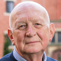
Nitric oxide, carbon monoxide and hydrogen sulfide: a microbial perspective of the trinity
Robert K. Poole
Emeritus Professor of Microbiology, The University of Sheffield, Sheffield, UKNitric oxide, carbon monoxide and hydrogen sulfide are very small molecules but of fundamental importance in all areas of biology and biochemistry. They are often called ‘gasotransmitters’, i.e. small, generally reactive, gaseous molecules that, in solution, may be generated endogenously in an organism and exert important signalling roles. However, this definition conceals their important effects on microbes, namely as poisons, participants in redox chemistry and as small ligands, notably to haems. I will examine whether the term ‘gasotransmitter’ is really appropriate and give an overview of these molecules, drawing particular attention to common misperceptions, recent studies and controversies that may cloud our understanding and study of these molecules.
Robert K. Poole
Robert Poole graduated with a First Class Honours degree in Microbiology and obtained his PhD and DSc (a higher doctorate) degrees in the University of Wales. After a postdoctoral period in The University of Dundee, he has held professorial roles at King’s College London and The University of Sheffield, where he was appointed West Riding Professor of Microbiology in 1996. His research interests have always focused on microbial (yeast and bacterial) respiratory systems, especially terminal oxidases and haemoglobins, and their interactions with O2, NO and CO. He has spent long periods of research at The Johnson Research Foundation in Philadelphia with Dr Britton Chance, Kyoto University and Cornell University. He is a long-standing Editor of the Journal of Biological Chemistry and of Advances in Microbial Physiology. He has an h-index of 64.
-
Seminar #10 - 18 November
Pharmacological perspectives from investigation of the gasotransmitter hydrogen sulfide in Down syndrome and Alzheimer's disease
15:00-15:10
Welcome and Introduction
Alessandro Giuffre
(Consiglio Nazionale delle Ricerche, Italy)15:10-15:50
The CBS/H2S system: a re-emerging therapeutic target in Down syndrome
Csaba Szabo
(University of Fribourg, Switzerland)15:50-16:30
Protective role of Sulfhydration in Alzheimer’s disease

Bindu D. Paul
(Johns Hopkins University, Baltimore, MD, USA)16:30-17:00
Questions and Answers
The CBS/H2S system: a re-emerging therapeutic target in Down syndrome
Csaba Szabo
Chair of Pharmacology, University of Fribourg, SwitzerlandDown syndrome (DS) is associated with significant perturbances in many morphological and biochemical features. Cystathionine-beta-synthase (CBS) is one of the key mammalian enzymes that is responsible for the biological production of the gaseous transmitter hydrogen sulfide (H2S). When H2S is overproduced, it can exert detrimental cellular effects, in part due to inhibition of mitochondrial Complex IV activity. An increased expression of CBS and the consequent overproduction of H2S is well documented in individuals with DS. Two decades ago, it has been proposed that a toxic overproduction of H2S importantly contributes to the metabolic and neurological deficits associated with DS. However, until recently, this hypothesis has not yet been tested experimentally. Recent data generated in human dermal fibroblasts show that DS cells overproduce H2S, which, in turn, suppresses mitochondrial Complex IV activity and impairs mitochondrial oxygen consumption and ATP generation. Therapeutic CBS inhibition lifts the tonic (and reversible) suppression of Complex IV: this results in the normalization of mitochondrial function in DS cells. H2S may also contribute to the cellular dysfunction via several other molecular mechanisms through interactions with various mitochondrial and extramitochondrial molecular targets. The current lecture provides a historical background of the field, summarizes the recently published data and their potential implications and outlines potential translational approaches (such as CBS inhibition and H2S neutralization) and future experimental studies in this re-emerging field of pathobiochemistry.
Csaba Szabo
Professor Szabo is an internationally recognized expert in the fields of oxidative and nitrosative stress, gaseous transmitters, cell death, cell dysfunction, cardiovascular, and inflammatory mechanisms. In the mid 90's he has pioneered the concept that identified the pathogenetic role of the nuclear enzyme PARP in promoting cell necrosis, and demonstrated its pathophysiological role in critical illness, cardiovascular and inflammatory diseases. His work in this area led to novel drug candidates that have progressed into clinical trials. For the last decade, much of his work focused on the biology of the novel endogenous gaseous mediator hydrogen sulfide (H2S); he discovered multiple novel roles of H2S in circulatory shock, reperfusion injury, angiogenesis, cancer and Down syndrome. This work has led to the identification of several novel therapeutic concepts based either on H2S donation or H2S biosynthesis inhibition.
Originally trained as a M.D/Ph.D. in Budapest, Hungary, he conducted a postdoctoral fellowship with Nobel Laureate Sir John Vane at the William Harvey Research Institute in London, England in the early 90’s. Between 1994 and 2018, as a professor of Pharmacology, Experimental Surgery and Anesthesiology, he has founded and directed several academic and biotechnology research groups in the USA in Ohio, Massachusetts, New Jersey, Washington and Texas. In 2018 he returned to Europe, where he serves as the Chair of the Department of Pharmacology at the University of Fribourg, Switzerland. He leads a research group that investigates the biological roles of PARP and H2S in health and disease, through employing the combined approaches of pharmacology, physiology, pathophysiology, cell biology, molecular biology, medicinal chemistry and translational science.
With a H-index of 136 and over 70,000 literature citations, he has been in the top 10 most cited pharmacologists in the world for the last decade. Professor Szabo has received numerous awards, including the Novartis Award of the British Pharmacological Society, the Dennis Gabor Innovation Award, the Texas Star Award, the Pharmacia-ASPET Award for Experimental Therapeutics and the John Vane Medal. He serves on the Editorial Board of numerous leading journals, including the British Journal of Pharmacology, Pharmacological Research, Nitric Oxide and Molecular Medicine. He is an Elected Fellow of the British Pharmacological Society and an Elected Member of the American Society for Clinical Investigation.Protective role of Sulfhydration in Alzheimer’s disease
Bindu D. Paul1,2,3
1Department of Pharmacology and Molecular Sciences, Johns Hopkins University School of Medicine,
2Department of Psychiatry and Behavioral Sciences, Johns Hopkins University School of Medicine,
3The Solomon H. Snyder Department of Neuroscience, Johns Hopkins University School of Medicine, Baltimore, MD, USAAlzheimer's disease (AD) is a progressive neurodegenerative disease that causes debilitating dementia. Pathological features of AD include deposition of amyloid plaques, neurofibrillary tangles (NFTs) and paired helical filaments in the brain. Mutations in the microtubule-associated protein Tau, a component of the NFTs, cause its hyperphosphorylation and aggregation in AD. Here we show that cystathionine γ-lyase (CSE), the biosynthetic enzyme for the gaseous messenger molecule, hydrogen sulfide (H2S), binds wild type Tau, which enhances its catalytic activity. By contrast, CSE fails to bind Tau P301L, a mutant that is present in the 3xTg-AD mouse model of AD. We further demonstrate that signaling by H2S is dysregulated in AD. H2S signals via a posttranslational modification termed sulfhydration/persulfidation, wherein –SH groups of reactive cysteine residues are converted to –SSH groups. Sulfhydration regulates several physiological processes which include vasorelaxation, response to inflammation and antioxidant defense mechanisms. We show that sulfhydration is diminished in 3xTg-AD mice as well as in human AD brains. Furthermore, H2S prevents hyperphosphorylation of Tau by sulfhydrating its kinase, glycogen synthase kinase 3β (GSK3β) and inhibiting its catalytic activity. Most importantly, we demonstrate that sulfhydration is diminished in AD, and administering the H2S donor sodium GYY4137 (NaGYY) to 3xTg-AD mice ameliorates motor and cognitive deficits in AD. Aging is a risk for developing AD and we further show that H2S signaling and sulfhydration is compromised during aging, which leads to impaired stress responses.
References:
Giovinazzo D, Bursac B, Sbodio JI, Nalluru S, Snowman AM, Whiteman M, Filipovic MR, Snyder SH* and Paul BD*. Hydrogen sulfide mediates neuroprotection in Alzheimer's disease by sulfhydration of GSK3β and inhibition of Tau hyper-phosphorylation. Proc Natl Acad Sci USA. 2021. Jan 26;118(4):e2017225118. doi: 10.1073/pnas.2017225118. (*Corresponding).
Paul BD. Neuroprotective Roles of the Reverse Transsulfuration Pathway in Alzheimer's Disease. Front Aging Neurosci. 2021 Mar 16;13:659402. doi: 10.3389/fnagi.2021.659402.Bindu D. Paul
Dr. Bindu Paul is an Assistant Professor of Pharmacology, Neuroscience and Psychiatry at the Johns Hopkins University School of Medicine, Baltimore, MD, USA. She obtained her PhD in Microbiology and Cell Biology from the Indian Institute of Science, Bangalore, where she studied the late gene transcriptional activator protein. C, from bacteriophage Mu with Dr. V. Nagaraja. Her post-doctoral studies involved the elucidation of gene regulatory processes modulated by the thyroid hormone at the National Institutes of Health, Bethesda, USA with Dr. Yun-Bo Shi. She discovered an essential role for steroid receptor coactivators in the context of thyroid hormone signaling during amphibian metamorphosis. Next, she analyzed signaling cascades modulated by the gaseous signaling molecules, hydrogen sulfide and nitric oxide, in the context of aging and neurodegenerative diseases such as Alzheimer’s disease and Huntington’s disease with Dr. Solomon Snyder (Sol) at the Johns Hopkins University, Baltimore, USA. For her contributions to the field, she received the Herbert Tabor Young Investigator Award from the American Society for Biochemistry and Molecular Biology in 2015. She continues her studies on redox active molecules and cysteine metabolism in the brain with a goal to develop novel therapeutics.
-
Seminar #11 - 2 December
Metabolic ROS production and resolution of inflammation: new perspectives for anticancer treatments
15:00-15:10
Welcome and Introduction
Mauro Maccarrone
(University of L'Aquila, Italy)15:10-15:40
Modulation of cancer cell proliferation by interfering with metabolic ROS production: new perspective for anticancer treatments
Maria Rosa Ciriolo
(Tor Vergata University of Rome, Italy)15:40-16:10
Proresolving lipid mediators as novel endogenous modulators of oxidative stress
Alessandro Leuti
(Campus Bio-Medico University of Rome, Italy)16:10-17:00
Questions and Answers
Modulation of cancer cell proliferation by interfering with metabolic ROS production: new perspective for anticancer treatments
Maria Rosa Ciriolo
Tor Vergata University of Rome, ItalyMetabolic plasticity enables cancer cells to efficiently switch their metabolic phenotypes between glycolysis and oxidative phosphorylation. Moreover, cancer cells can use aerobic glycolysis for rapid ATP production and biomass synthesis to facilitate tumorigenesis and modulate their metastatic potential. However, it is still largely unknown how cancer cells orchestrate gene regulation to balance their glycolysis and oxidative phosphorylation activities.
Here I will discuss recent data that shed light on how tumor cells adapt their metabolic pathways under forced fatty acid utilization or Krebs cycle modulation and highlight the importance of mitochondrial ROS signalling in this process, a pathway that might be amenable to therapeutic targeting.Maria Rosa Ciriolo
Full Professor and Coordinator of the PhD school in Cellular and Molecular Biology at University of Rome “Tor Vergata”, Rome, Italy. Head of the laboratory of “Biochemistry of Ageing” at IRCCS San Raffaele, Rome, Italy, Head of the redox Biochemistry group, that actively and productively works in the field of redox-mediated cell signaling downstream of ROS and NOS-mediated processes characterizing cancer, neurodegeneration and aging. These studies highlighted for the first time that a remarkable number of proteins can fit a double role in signaling by sensing alterations of the cellular redox state by means of reactive cysteine residues. Part of the research focused largely on the molecular mechanism linking copper metabolism and SOD1 in the pathophysiology of neuronal cell death.
She is author of more than 200 papers on high-ranking journals; 20 book chapters and 110 abstracts of International and National Congresses. H-index 60, Total citations >17000.Proresolving lipid mediators as novel endogenous modulators of oxidative stress
Alessandro Leuti
Campus Bio-Medico University of Rome, ItalySpecialized proresolving mediators (SPMs) are a novel class of endogenous lipids, derived by ω-6 and ω-3 essential polyunsaturated fatty acids such as arachidonic acid (AA), docosahexaenoic acid (DHA), and eicosapentaenoic acid (EPA) that trigger and orchestrate the resolution of inflammation, which is the series of cellular and molecular events that leads to spontaneous regression of inflammatory processes and restores tissue homeostasis. These lipids are emerging as highly effective therapeutic agents that exert their immunoregulatory activity by activating the pro-resolving pathway, as reported by a consistent bulk of evidences gathered in the last two decades since their discovery. The production of reactive oxygen (ROS) and nitrogen (RNS) species by immune cells plays indeed an important role in the inflammatory mechanisms of host defence, and it is now clear that oxidative stress, viewed as an imbalance between such species and their elimination, can lead to many chronic inflammatory diseases. The main role of SPMs is confining the inflammatory response to avoid its aberrant prolongation, which they achieve by directly acting on the activation of both innate and adaptive immune cells; however, in recent years, they are also emerging as modulators of oxidative stress, a task that they achieve by either inhibiting the molecular pathways that these molecules trigger, or by inducing the expression of endogenous antioxidant systems. This property is of particular relevance in the genesis and clinical course of a number of chronic disorders, and suggest that SPMs might represent a valuable target in understanding inflammatory diseases in which the production of ROS and RNS – or the impairment of the antioxidant machinery – represent a main pathogenetic mechanism.
Alessandro Leuti
Dr Alessandro Leuti is assistant professor in biochemistry at Campus Bio-Medico University of Rome, where he works at the Biochemistry and Molecular Biology Unit. His main focus of research is the role of endogenous bioactive lipids as immune modulators in inflammatory pathologies, with particular attention at ω-3- and ω-6-derived specialized pro-resolving mediators (i.e., resolvins, protectins, maresins and lipoxins). He is been author or co-author in more than 20 peer-reviewed scientific papers (h-index: 13).
-
Seminar #12 - 16 December
Aberrant redox responses in neurodevelopmental conditions: the case of autism and Rett Syndrome
15:00-15:10
Welcome and Introduction
Luciano Saso
(Faculty of Pharmacy and Medicine, Sapienza University of Rome)15:10-15:30
OxInflammation in Rett Syndrome
Giuseppe Valacchi
(Department of Neuroscience and Rehabilitation, University of Ferrara, Italy)15:30-15:50
Deciphering the role of cerebral redox imbalance in Rett Syndrome

Michael Müller
(University Medical Center Göttingen, Inst. Neuro- and Sensory Physiology, Göttingen, Germany)15:50-16:10
Mitochondrial impairment in Autism Spectrum Disorders
Alessandra Pecorelli
(North Carolina State University, Animal Science Dept., Plants for Human Health Institute, Kannapolis, NC, USA)16:10-17:00
Questions and Answers
OxInflammation in Rett Syndrome
Giuseppe Valacchi
Department of Neuroscience and Rehabilitation, University of Ferrara, ItalyRett Syndrome (RTT) is a progressive neurodevelopmental disorder resulting from mutations in the X-linked gene encoding methyl-CpG-binding protein 2 (MeCP2). With an estimated incidence of one case per 10,000 live births, RTT is the second most commonly recognized genetic cause of mental retardation in females. Although brain is the main organ affected by MECP2 mutation, RTT manifest a complex multisystem nature with a wide range of clinical manifestations. Among a different variety of metabolic alterations, an oxinflammatory condition characterized by the positive feedback loop between unbalanced redox homeostasis and atypical immune function appears to be a critical player in the onset and progression of RTT pathophysiology. Oxinflammatory biomarkers show a strong correlation with RTT clinical stage and severity in brain and periphery of both animal models and RTT patients. Investigations on the underlying molecular mechanisms reveal an impaired enzymatic defensive activity coupled with an uncontrolled activation of NADPH oxidase in RTT patients' cells. Moreover, alterations in mitochondrial bioenergetics, dynamics and quality control pathways by mitophagy link the redox imbalance to immune dysfunction, by promoting the accumulation of damaged mitochondria able to induce an aberrant NLRP3 inflammasome signalling. In addition, increased levels of lipid oxidation end-products such as 4-HNE and HODEs, oxidized arachidonic and linoleic acid metabolites respectively, can affect RTT immune responses as indicated by the proinflammatoty plasma milieu together with morphological abnormalities of immune cells. Altogether, our data provide new insights in elucidating the altered biological processes associated with the oxinflammation phenomenon in RTT and, furthermore, can help in identify new potential therapeutic targets for a disorder that, at today, is still orphan of therapies.
Giuseppe Valacchi
Giuseppe Valacchi obtained his B.S. with Laude in physiology, MS in physiopathology and his PhD degree in Cell Physiology and Neuroimmuno-physiology at the University of Siena. During his training he has worked as “exchange PhD student" in the “Department of Molecular and Cell Biology at the University of California at Berkeley" where he started out his studies on the effect of environmental oxidative stress such as O3 and UV on skin physiology. After his PhD graduation, he continued working as Post Doc at the University of Berkeley, California (USA) then in the “Department of Internal Medicine" and in the "Department of Nutrition" at the University of California at Davis (UCD). In 2005 he become Faculty and member of the Academic Federation in the Department of Internal Medicine (UCD). At the end of 2006 he was awarded with the “progetto rientro dei cervelli”, granted by the Italian Ministry of Health and returned to Italy at the University of Siena as Assistant Professor until 2011. In 2011 he was appointed as Associate Professor in Physiology at the University of Ferrara and in 2021 was promoted to Full Professor in Physiology. From 2008 is also Adjunct Prof. at Kyung Hee University, Seoul, South Korea and starting from August 2016 he was first Associate Professor and then Full Professor in Regenerative Medicine at North Caroline State University. His research has been focused in understanding the cellular, and molecular mechanisms that define the tissues physio-pathological responses to altered redox homeostasis with special focus on cutaneous tissue. He is a member of the SFRR Europe Council since 2017 and he is author of more than 280 peer reviewed internationals papers (most of them related to the effect of pollution in target organs), 15 book chapters, one book. He has been invited speaker to more than 100 international conferences and organizer/Chair to over 50 international meetings. He has won several awards among them the Entelligence Award from Actelion, OCC Young Investigator Award, Science and Education Award and recently, in 2019, the Exposome grant. In 2018 he was awarded with the “Doctorate Honoris Causa” in Biochemistry and Pharmacy from the University of Buenos Aires (rank n. 72 among the over 3,000 universities in the world) for his work in the redox biology field, in addition he is the recipient of the 2020 Clinical Research award from the SFRR-E. He is the Associate Editor of several international journals among which is Mediators of Inflammation, Frontiers in Cellular Biochemistry, World Research Journal of Biochemistry, Journal of Complementary and Traditional Medicine, Biomed Research International (Dermatology Subjects), Oxidative Medicine and Cellular Longevity, Antioxidants; in addition he is member of the Editorial Board of several journals including Genes and Nutrition, Open Biochemistry Journal, Archives in Biophysics and Biochemistry, Clinical Immunology Endocrine & Metabolic Drugs, Frontiers in Inflammation Pharmacology, Clinical Anti-Inflammatory & Anti-Allergy Drugs, BioFactors, Cosmetics, Free Radicals in Biology and Medicine.
Deciphering the role of cerebral redox imbalance in Rett Syndrome
Michael Müller
University Medical Center Göttingen, Inst. Neuro- and Sensory Physiology, Göttingen, GermanyRett syndrome (RTT) is associated with early mitochondrial dysfunction and systemic oxidative stress. Mitochondria of MeCP2-deficient (Mecp2-/y) mouse hippocampus seem partly uncoupled, apparently consume higher rates of oxygen, and release increased amounts of reactive oxygen species (ROS). In concert with less efficient cell-endogenous scavenging systems, this culminates in cellular redox dysregulation. As these alterations manifest already in presymptomatic mice, redox imbalance and the associated oxidative stress are considered potential mechanisms facilitating disease progression. The beneficial effects of antioxidant-based therapies support this scenario. To decipher the molecular details of this redox impairment, we used the genetically-encoded optical redox sensor reduction-oxidation sensitive green fluorescent protein (roGFP) and performed excitation-ratiometric fluorescence imaging in hippocampal cultures. Quantitative redox-imaging confirmed an oxidative burden in cultured neurons and astrocytes of neonatal Mecp2-/y hippocampus. Taking a closer look at neurons revealed that the cellular redox-imbalance affects cytosol and mitochondria. In organotypic hippocampal slices, acute redox challenge by H2O2 and severe hypoxia elicited intensified oxidizing and reducing transients in Mecp2-/y neurons, respectively. Also neuronal stimulation by various neurotransmitters evoked intensified oxidizing shifts in the cytosol of Mecp2-/y neurons, suggesting that even physiological stimuli are sufficient to provoke overshooting redox responses. In mitochondria, neurotransmitter-evoked redox changes were more moderate than in cytosol, and the genotypic differences were less pronounced. This identifies the cytosol as an important ROS source and as a less stably redox-buffered compartment. As all of these redox changes were already evident in tissue of neonatal and presymptomatic mice, it will be crucial now to map these changes as RTT emerges and slowly progresses in severity. The ideal tool for this future endeavour are our recently generated roGFP-Rett mice, which stably express the roGFP redox sensor in cytosol and mitochondria of central neurons. These mice will now enable a brain-wide redox mapping to identify the particular brain areas affected. Also, they will allow for the first time to correlate neuronal redox alterations with disease progression as well as symptom severity, and finally will prove pivotal for verifying the efficiency of redox-stabilizing therapeutic approaches in RTT.
Supported by: Cluster of Excellence and DFG Research Center for Nanomicroscopy and Molecular Physiology of the Brain (CNMPB), University Medical Center Göttingen, State of Lower Saxony, International Rett Syndrome Foundation (IRSF), Deutsche Forschungsgemeinschaft (DFG, grant #456368804).Michael Müller
Michael Müller is an Associate Professor (Apl. Professor) in the Institute of Neuro- and Sensory Physiology at the University Medical Center Göttingen. He received his Diploma in Biology (1992) and his PhD in Neurosciences (1996) from the Heinrich-Heine-University of Düsseldorf (Germany). As a Research Associate at the Department of Cell Biology at Duke University Medical Center (Durham, NC, USA, 1997-1999), he started focusing at the responses of neuronal networks to hypoxia. Being appointed a Junior-Professor of Molecular Neurophysiology at the University Medical Center Göttingen in 2004, he established an independent research group to study the modulation of central neurons by metabolic compromise and by redox-related signals. From 2009 - 2018, Dr. Müller has been a board member of the former DFG Research Center and Cluster of Excellence “Nanomicroscopy and Molecular Physiology of the Brain (CNMPB)” and one of the elected scientific coordinators of the research field “Neurodevelopmental Disorders: From Synaptopathies to System Dysfunction”. Over the past 10 years, Dr. Müller´s research team has increasingly focused on neuronal network dysfunction, mitochondrial impairment, and redox alterations in mouse models of Rett syndrome. By establishing the quantitative imaging of cellular redox-dynamics, his group made important contributions to decipher those mechanisms underlying the oxidative stress in Rett syndrome and to clarify the potential merits of antioxidant- and free radical scavenger-based pharmacotherapies.
Mitochondrial impairment in Autism Spectrum Disorders
Alessandra Pecorelli
North Carolina State University, Animal Science Dept, Plants for Human Health Institute, Kannapolis, NC, USAAutism Spectrum Disorder (ASD) is a multifactorial condition caused by the interaction between genetic predisposition and increased vulnerability to a variety of external perturbations in prenatal and perinatal periods. However, the pathogenic mechanisms underlying the development of ASD still remain elusive. Based on growing evidence, ASD pathophysiology appears clearly affected by the oxinflammation phenomenon, i.e. a harmful interplay between disturbed redox homeostasis and impaired immune response, with mitochondrial dysfunction as central hub between them. To investigate more in detail the altered mitochondrial function in this pathology, here, we analyzed primary fibroblasts isolated from ASD patients by measuring mitochondrial bioenergetics, ultra-structural and dynamic parameters. In addition, as mitochondria and inflammasomes are closely related to each other, we also evaluated components and function of the inflammasome machinery. High levels of 4-hydroxynonenal (4-HNE) protein adducts together with increased NADPH oxidase (NOX) activity and mitochondrial superoxide production coupled with a compromised antioxidant response guided by a defective Nuclear Factor Erythroid 2-Related Factor 2 (Nrf2) pathway confirmed an unbalanced redox homeostasis in ASD. Moreover, ASD fibroblasts showed overactive mitochondrial bioenergetics associated with atypical morphology and altered expression of mitochondrial electron transport chain (ETC) complexes and dynamics-regulating factors. Furthermore, priming with lipopolysaccharide (LPS) followed by stimulation with the danger signal adenosine triphosphate (ATP) induced a significant increase in the levels of several inflammasome constituents including ASC, caspase-1 and interleukin-1β in ASD cells. Overall, our data suggest a new possible pathogenic mechanism underlying ASD based on the dysfunctional interaction between redox and immune responses, as indicated by the concomitant presence of mitochondrial defects and inflammasome activation in cells from patients affected by this complex neurodevelopmental disorder.
Alessandra Pecorelli
Alessandra Pecorelli obtained her PharmD degree cum laude in 2004 and her Ph.D. degree in Cell Biology and Pathophysiology in 2013 at the University of Siena (Siena, Italy). Between 2005 and 2009, she was Postdoc at the Department of Physiology in the University of Siena. Since that time, her primary research interests are focused on the role and contribution of oxidative stress and inflammation in several pathophysiological conditions, focusing first in the effects of exogenous stressors on target tissues, such as lung and skin. During the PhD course at the Department of Molecular and Developmental Medicine (University of Siena), she started to study the pathophysiological mechanisms involved in neurodevelopmental disorders such as Autism Spectrum Disorders and Rett syndrome. After completing her doctoral degree, she continued her studies in Rett and Autism at the same university and at the University of Ferrara as Postdoc. Since the end of 2016, she moved to the United States at North Carolina State University where she works as Senior Postdoctoral Research Fellow at the Plants for Human Health Institute in the Research Campus in Kannapolis (NC, USA). Following her scientific work, first in Siena and Ferrara, and now in USA, she has become a prominent expert in the field of Rett and Autism. With more than 100 published articles, of which over 40 on Rett Syndrome and Autism Spectrum Disorders, she is now recognized as one of the leading young experts in the biochemical mechanisms involved in these diseases. She participated in several international conferences and her work has been awarded several times. Of note are her prizes. She has won three “Young Investigator Award” by OCC (Oxygen Club of California). In 2011, at the “Second International Conference on Environmental Stressors in Biology and Medicine” in Siena, Italy (5-7 October 2011); in 2015, at the World Congress "Oxidants and Antioxidants in Biology" in Valencia, Spain (24-26 June 2015); in 2021, at the “20th Biennial Meeting for the Society for Free Radical Research International (SFRRI 2021)”, Virtual Conference (15-18 March 2021). In 2019, she has received the prestigious “Catherine Pasquier Award" from the Society for Free Radical Research – Europe at the SFRRE Meeting “Redox Homeostasis: from Signaling to Damage” in Ferrara, Italy (19-21 June 2019).